Since their inception, opinions over the value of high-volume public programmes for breast screening have diverged, and there is currently much controversy in the press about their effectiveness. Regardless of which side one takes in the debate, there is certainly a growing emphasis on finding new ways to conduct these large public health programmes.
In many European countries women are encouraged to have regular screenings using low-energy X-rays in order to detect masses or micro-calcifications early. Women in the UK, for instance, are invited to have mammograms every three years after the age of 50, and the programme is gradually being extended to include women between 47 and 50, although this is currently under review. It is widely believed that these programmes help reduce the number of deaths from breast cancer, but there are those who disagree.
Heated controversy
Recently published research by Peter Gøtzsche, director of the Nordic Cochrane Collaboration, is the result of over ten years of investigation and analysis of data from breast screening trials. It calls into question the viability of high-volume, population-based screening programmes on the basis that they harm more women than they save.
Gøtzsche suggests that the harm comes from overdiagnosis and invasive treatment, while relatively few lives are saved. His strongly worded report accuses some in the medical profession of misconduct, suggesting that evidence has been buried in order to sustain public mammography programmes.
Furthermore, he calls into question the commonly quoted statistic that regular screening cuts breast cancer deaths by 30%, suggesting that it saves one life for every 2,000 women screened, but harms ten others, often through the treatment of cancerous cells that would disappear or never develop into the disease. He claims that cancerous cells that would pose no long-term problem are often removed through surgery that in 60% of cases results in women losing a breast.
His views certainly run contrary to the majority of experts in the field, and represent the extreme end of the criticism of public breast screening programmes. Nevertheless, they do highlight problems that are well understood and have led to the ongoing search for alternative methods of conducting high-volume screening programmes. The 2D imaging used in traditional mammograms does have its limitations.

US Tariffs are shifting - will you react or anticipate?
Don’t let policy changes catch you off guard. Stay proactive with real-time data and expert analysis.
By GlobalData"There are problems with traditional mammography for women with dense breast tissue, and young women in particular. There is no doubt that there are challenges concerning false positives, which in Europe are at the level of around 3%, and overdiagnosis, although we don’t have accurate figures for how many diagnosed cancers would never have presented clinically. There are huge differences of opinion on that, with figures ranging from 3-50%. The higher figures Gøtzsche cites are in contrast with most experts’," says Professor Dr med. Per Skaane of the department of radiology and nuclear medicine at Oslo’s Ullevaal University Hospital.
"For the last decade we have been discussing how to improve the screening process with something other than traditional mammography. There are clinics that offer personalised screening using MRI scans or hand-held ultrasound (HHUS), but these are low-volume programmes and these techniques cannot be included in high-volume programmes because of the time and expense they require. MRI, for example, is too expensive except for high-risk women," he adds.
The candidates
Skaane sees two possible candidates to replace, or complement, traditional mammography: automated breast volume scanning (ABVS) and digital breast tomosynthesis. Ultrasound of the whole breast has so far suggested higher rates of cancer detection are possible when used in conjunction with mammography for women with dense breasts or higher risk factors. Obviously, this is not yet a replacement technology, but a complementary one, and it incurs a higher cost. There are also other hurdles for automated ultrasound to overcome. Hand-held ultrasonography is done by a radiologist, and studies from the US show that it takes 19 minutes a case, which is too slow for high-volume screening programmes. Mammographic interpretation by radiologists takes only 50 seconds a case. Automated ultrasound could be the answer since it requires less radiologist time. In Europe, only Austria is looking at ultrasound as an adjunct to mammography for the time being. "Tomosynthesis is the only other option," observes Skaane.
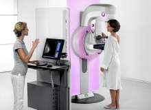
Digital tomosynthesis creates a 3D image of the breast using X-rays to reveal its inner architecture without superposition of disturbing overlaying tissue. Better visualisation of the breast tissue based on 1mm thick slices offers the potential for more detailed examination of the breast parenchyma compared with ultrasound.
"The first clinical results show increased specificity, so there are fewer false positives, but those are results from the US, where the callback rate is much higher – around 10-12%, compared with Europe, where it is only 3-5%. So, tomosynthesis may be of less importance in this regard in Europe. For it to work in high-volume screening programmes in Europe it must increase the sensitivity, but there are no studies worldwide so far that have shown an increased cancer detection rate by tomosynthesis.
"The exciting thing is that tomosynthesis is fast, and it takes only four seconds more to take the DBT by the radiographers and about 85 seconds a case for the radiologists to do the interpretation, which is acceptable even though it is more time than it takes to interpret a 2D mammogram. But we don’t know yet if the results make tomosynthesis viable," he adds.
Skaane is running a two-year study, which will report its first year results soon, to understand the role tomosynthesis could play in high-volume screening. Women between 50 and 69 who use the screening facilities at Oslo University Hospital are offered an additional 3D scan on their scheduled visit. As many as 20,000 women will have participated in the study at the hospital, which screens up to 80 women each day. A similar study is under way in Malmo, Sweden.
"The technology can only be discussed seriously once we have the results from prospective trials in a screening setting. We know that the radiation dose is a challenge. The interpretation time and the hardware cost are acceptable. With ultrasound, the problem is that the decision time is too long. So, we need more results from trials and we need to understand how we can use tomosynthesis in a way that requires a lower dose of radiation," notes Skaane.
"Some women with healthy breasts could have just one mediolateral-oblique (MLO) image rather than two. For women with very dense breasts we could do one MLO and one automated ultrasound, and we could use tomosynthesis for breasts of medium density. But we need prospective trials to investigate this," he adds.
Combining screening technologies
For tomosynthesis, the relatively short time required to interpret the images is certainly one factor that is in its favour. In the study at Oslo University Hospital, four radiologists interpret each digital image, and there are independent readings of conventional 2D mammograms, 2D plus computer-aided detection (CAD), 2D plus tomosynthesis and synthetic 2D – reconstructed 2D images based on the 3D dataset – plus tomosynthesis.
Using different combinations of imaging technology allows a comparison of their effectiveness, highlighting the strengths of each. The use of 2D and 3D imaging brings a number of advantages. The combination of synthetic 2D and tomosynthesis is of interest since synthetic 2D has the ability to highlight microcalcifications and 3D (tomosynthesis) to reveal small spiculated masses and distortions.
With this more detailed view of the breast tissue, it should be possible to highlight a greater number of cancers at an early stage in their development, but confirmation of this will only be possible when the results of the full study are examined in detail.
Previous studies’ results comparing the specificity of 2D mammography and tomosynthesis show the latter to have superior performance. When it comes to sensitivity, especially the detection of small spiculated masses and distortions, Skaane believes that tomosynthesis may well have an important role to play alongside 2D imaging in the context of high-volume screening programmes. After all, the main goal is to detect more cancers.
The higher specificity of tomosynthesis could be of particular benefit in the US, where callback rates are much higher than in Europe. In the European setting with much lower recall rates, increased sensitivity with detection of more cancers would be given more priority. Yet, there are still obstacles to overcome.
While it is likely that tomosynthesis will help to reveal more cancerous tissue at an earlier stage, a key focus for the future will be lowering the dose of radiation that is required to generate the 3D images.
"If we could reduce the radiation dose to make it comparable to today’s traditional mammograms then that would certainly be of interest to a lot of people. At the moment, tomosynthesis requires about the same radiation dose as mammography, so the combination of 2D and 3D would double the dose. This is not acceptable for a screening programme," comments Skaane.
For now, it seems, there will be no quick end to the controversy over how public programmes of breast screening are conducted, but all sides in the debate want to see further development of technologies that can yield more accurate results in a sufficiently rapid way. Work is underway to assess different systems and combinations of technologies, and every well-run trial contributes more useful data.
Depending on the results of studies like the one being run in Oslo, an improvement in the accuracy of high-volume breast cancer screening could be possible within a couple of years. Furthermore, progress on lowering the radiation dose to which women will be exposed would also be expected. Skaane is certainly optimistic about the future and believes that women will undergo safer screening procedures that will achieve a higher rate of accurate diagnosis thanks to the use of tomosynthesis.
"In my view, in high-volume screening, whatever technology we talk about must be acceptable in terms of the time per exam, so we can’t use techniques that need contrast screening, for example. We may have high-resolution CT scans in a couple of years but they can’t be used for high-volume screening programmes. Automated ultrasound and tomosynthesis are the only real options today. The challenge of false positives is a big one, as ultrasound easily results in many more, so we would have to be very careful before we implement it. Tomosynthesis does not have the same problem," he explains.
"Nevertheless, to justify the examination time and the radiation dose, tomosynthesis must pick up significantly more cancers. If it does not, then there is no place for it in organised European screening programmes. It is too early to tell from the results of our study, although it certainly does seem to find more. But the detection rate needs to be significantly higher than mammography, and that is something we don’t know yet."

This article was first published in our sister publication Medical Imaging Technology.


