Cardiovascular disease (CVD) is a leading cause of death and disability worldwide. According to the American Heart Association, CVD claims more lives in the US than cancer, respiratory disease, accidents and diabetes combined.
CVD risk factors such as obesity and diabetes are also on the rise. These adverse health trends are being observed in developed countries as well as the growing middle classes of emerging economies such as China and India.

Discover B2B Marketing That Performs
Combine business intelligence and editorial excellence to reach engaged professionals across 36 leading media platforms.
The most prevailing cardiac disease is coronary heart disease (CHD), which involves the accumulation of atherosclerotic plaque within the walls of the coronary arteries, eventually blocking the supply of oxygen and nutrients to heart muscle.
Early detection and diagnosis is key for improving outcomes for any disease. Although cancers can be screened for, diagnosed and monitored by several imaging modalities, effective tools to do the same for CHD do not exist. Many patients only become aware that they have the condition after they suffer their first heart attack, if they survive it.
Treadmill stress tests and myocardial perfusion imaging provide limited sensitivity and specificity, so the primary diagnostic tool for CHD is X-ray angiography, which is carried out in a catheterisation laboratory and is typically only available to patients at an advanced stage of the disease. The procedure is only effective at detecting coronary arteries that are already blocked; it does not reveal the overall build-up, characterisation or distribution of plaque.
If significant stenoses (blocked arteries) are found, it is possible to proceed to a therapeutic stage in the same or a follow-up session, and relieve them using balloon angioplasty and stents.

US Tariffs are shifting - will you react or anticipate?
Don’t let policy changes catch you off guard. Stay proactive with real-time data and expert analysis.
By GlobalDataHowever, the procedure ends at the diagnostic stage for many patients. This could be because significant stenoses are not found, because a total occlusion is found that cannot be cured in catheterisation or because other conditions are found that warrant a bypass operation rather than balloon angioplasty.
It’s for these reasons that a non-invasive diagnostic device that can provide images of the coronary arteries and chart the distribution and characterisation of plaque is highly desired.
CT in cardiac imaging
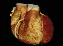
CT has not been traditionally used in cardiac imaging because the heart is constantly moving. This situation has changed in recent years with the development of 64-slice fast rotation CT that is almost able to ‘freeze’ heart motion, providing a viable non-invasive alternative to diagnostic catheterisation. Cardiac CT angiography only requires an intravenous injection of dye into the arm followed by a CT scan of the heart, and 64-slice CT can typically image the entire heart in six to eight seconds, or within ten heartbeats.
An ECG signal is used to gate data acquisition so that only a relatively narrow phase of the heart cycle is used for image reconstruction, the phase being selected in order to minimise motion artifacts.
Since the introduction of 64-slice CT in 2006, several more-advanced models have been introduced. One vendor offers a 320-slice CT that can capture the entire heart volume in one phase of a single heartbeat. Another vendor offers a dual-source CT that, combined with high-pitch spiral acquisition mode, can do the same. These and other advanced protocols not only improve temporal resolution and reduce blur, but reduce patient radiation exposure.
The main application of CT in cardiology today is as a ‘gatekeeper’ to the catheterisation laboratory for low and intermediate-risk patients known or suspected to suffer from CHD. A CT scan can reveal the overall deposition and composition of calcified and lipid-based plaque as well as stenoses. With cardiac CT angiography, there is no risk of vascular damage, heart attack or stroke; after the scan the patient can resume normal activities immediately.
According to some estimates, one in three people referred to cardiac catheterisation ultimately find they do not have any significantly blocked arteries, so using CT as a diagnostic stage can be an excellent way to reduce healthcare cost, avoid the risk of complications and eliminate the need for a hospital stay.
Most studies quote negative predictive values for the detection of clinically significant stenoses in the coronary arteries of 98-99% and higher, namely if the CT determines the arteries are not blocked, it is safe to assume they are indeed not blocked. But the positive predictive values are lower, depending on the equipment, protocol and patient population under study. Some of the issues affecting the positive predictive values are high or unstable heart rates and excessive accumulation of calcified plaque in the artery walls.
Another common application of CT in cardiology is the quantitative measurement of calcium load in the coronary arteries, referred to as the calcium score. This is a strong independent risk indicator for coronary events: high calcium score (Agaston score > 600) means an increased risk by a factor of 26 relative to a healthy condition (Agaston score = 0). Some experts believe that calcium score tests will eventually become a widely used screening test; however, such development is pending a reduction in the level of radiation dose.
Cardiac CT has been demonstrated to be effective for anatomic and functional imaging of the heart, wall-motion abnormalities, congenital heart disease, valve functionality and calcification, and bypass graft patency. It can also measure perfusion of the myocardium muscle, potentially replacing single photon emission CT as a primary diagnostic tool of myocardium physiology.
Repeated surveys among cardiologists have revealed high levels of interest in integrating cardiac CT into workflow, and around 4,000 have joined the recently founded Society for Cardiovascular Computed Tomography. Scores of cardiac CT courses and training programmes are offered to cardiologists, and a dedicated Journal of Cardiovascular Computed Tomography has been launched by Elsevier. Some cardiologists believe that Cardiac CT will become the ultimate differentiating tool for determining the treatment path for all low and intermediate-risk patients.
Dedicated cardiac CT versus whole-body CT
Although there is a growing interest in the use of CT as a part of cardiac disease management, there is no equipment on the market that is optimised specifically for this application.
To date, cardiac CT imaging is performed on the high-end and premium models of general purpose whole-body CT scanners adapted to acquire scan data in synchronisation with an ECG signal. Whole-body CT scanners have been instrumental in the development of cardiac CT and the creation of substantial clinical evidence in its support, but they have a number of inherited limitations that can be circumvented by using a system specifically designed for cardiac imaging.
1. Image quality
This is still unsatisfactory, resulting in equivocal test results. Most scanners require several seconds to image the heart, and volumetric images are obtained by stitching the data from different heartbeats. The heart does not exactly return to same position every beat so this stitching process generates blur and artifacts.
This can be avoided by a scanner that captures the entire heart within a single beat, a feature available today only on two top-priced scanners. Other image quality issues are related to cone beam reconstruction artifacts, sensitivity to arrhythmia, insufficient spatial resolution and high pulse rate.
2. Temporal resolution
Current CT scanners achieve high temporal resolution by fast mechanical rotation of the gantry. Whole-body CTs are limited in rotation speed as they carry heavy high power assemblies on the rotor, which are subject to immense centrifugal forces. A smaller diameter, light rotor would enable higher rotation speed and better freezing of heart motion.
3. Radiation dose
Typical dose levels for cardiac CT angiography studies are in the range of 2-15mSv, the main factors being the equipment, scan protocol and method of ECG gating. A limited field of view scanner, focused on the heart, with prospective ECG gating protocols that gate the radiation off during most of the heart cycle, can achieve the same image quality at routine radiation doses below 1mSv.
4. Imaging outside the heart
Whole-body CT inevitably irradiates the entire thorax and generates images of the chest alongside the heart. While this has a potential advantage of incidental findings, it involves unnecessary irradiation as well as workflow and liability issues, since the cardiologist who reads the heart images may not be qualified to interpret the images outside the heart. The issue is resolved with a cardiac dedicated scanner.
5. Cardiac workflow
Most whole-body scanners are installed in radiology departments and institutes, operated by radiologists and connected to the organisation’s radiology information system. From a cardiologist’s perspective, the cardiac imaging modalities should be on hand, operated according to the cardiac centre priorities and connected to the cardiology information system. This can be readily achieved with a dedicated machine.
6. Size, weight and power
Because whole-body scanners have to handle a variety of patients, body parts, scan conditions and protocols, the machines are heavy, require a large amount of floor space and consume a lot of power, which are not readily available in many facilities. There are substantial costs involved in site preparation. A dedicated cardiac CT can avoid this.
7. Cost
Although the price of 64-slice CTs is falling, the more advanced premium-tier scanners are inherently expensive to purchase and operate, affecting the cost of the test, profit margins and the cost of disease management. A dedicated cardiac CT can provide cardiac imaging performance exceeding that of the most advanced whole-body scanners, while keeping the price tag far lower.
Arineta cardiovascular CT scanner
Arineta is an Israel-based start-up company with a mission to provide innovative imaging solutions for improved cardiac care.
The company has developed a proprietary, novel-design CT scanner that is based on a new architecture and optimised for cardiovascular imaging. The Arineta design drives down costs by eliminating aspects that are required for whole-body imaging but are irrelevant for cardiac scanning, while optimising operation for a range of cardiac imaging procedures. The design is covered by pending patents.
The Arineta product will be a compact, high-performance imaging system that provides high-resolution, motion-free, artifact-free, volumetric images of the heart. The product addresses the specific requirements of cardiology procedures and the need for enhanced information during the diagnostic, interventional, and follow-up stages.
In addition to imaging of the heart, the Arineta product will be optimised for other cardiovascular studies, including those of the aorta, the carotid artery and the renal arteries.
Compared with conventional whole-body CT scanners, the Arineta device:
- provides imaging of the entire heart in a single heartbeat
- has coverage equivalent to a 330-slice CT scanner
- eliminates cone beam artifacts
- has improved spatial and temporal resolution
- has a limited field of view focused on the heart, not on the entire thorax
- has reduced radiation and contrast dye dose
- can be highly integrated in cardiology workflow
- is approximately a third of the weight and requires half of the floor space
- is substantially less expensive to own and operate.
The Arineta scanner is still under development and is not yet available for clinical work.


